Answer above (read it if you haven’t been to this one before).
OK, since I will not play in the outrage of the week fan club, perhaps I can interest all three of you in “guess the diagnosis”. I may never do this again, and may do it every week. The winner gets pride in smugly affirming their intelligence, everyone else gets to know the diagnosis. All diagnoses are made by me, and I’m also the judge, so appeals will be brief.
Adult male, brought by EMS after an MVA. Complains of Left chest pain and severe shortness of breath. A chest xray is obtained:
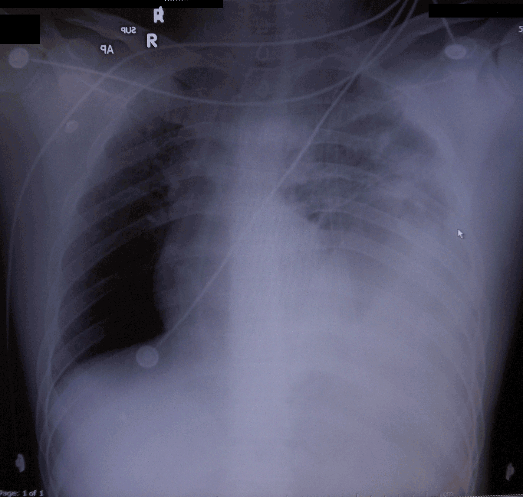
Supine, Portable AP CXR
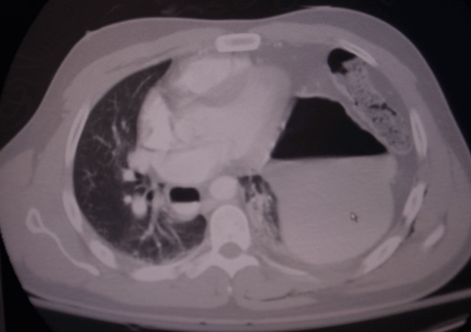
OK, have a good look, then decide (that’s how it works in the real world). A CT is below to confirm or refute your diagnosis.
A CT of the chest with IV, but without oral contrast:
OK, it’s not much of a contest, but it beats what I’ve been doing.
Contest closes 5-19, and I’ll post the correct diagnosis, and things I learned from it.

Rupture of the left hemidiaphragm. CT shows stomach in the posterior aspect of the left pulmonary cavity with atelectasis of the left lung lateral and anterior on the CT.
Duh, he’s got tons of wires running through his body!
… I was gonna say pneumo, but Tim’s probably right.
Oh. MVA = Motor Vehicle Accident. Yeah, probably more trauma related than a random pneumo. Whoops.
(History makes 80% of the diagnosis!)
Well I’m not a medical person by any stretch of the imagination but it seems that the left side is much darker than the right (I assume that we are supposed to look somewhat symetrical). So since I’m guessing that MVA is a car accident, my guess is that something got pushed (spleen, liver, something like that) into the lung area. Maybe it was one of those stupid steering wheel trays that I have seen some people use WHILE driving :-)
Left sided Hemothorax.
X-ray looks like there is a frac of the 4th rib on the left side, but I could be wrong,
I think the opacity is too diffuse to be …..
…..maybe not.
The CT shows the left side of the abdominal contents displaced superiorly. I can see stomach, and what I am guessing is splenic flexure of the colon. Spleen is UA.
I guess that Tim Sturgill is right, I mean I am just a lowly med student.
I would want to auscultate anyway. I figure that if I hear bowel sounds in the left thorax, then I guess we know.
I am assuming this is an upright PA view. Left hemothorax with communited fracture of the left 5th rib. Status-post chest tube thoracotomy on left. I don’t see an air-fluid level within the left hemithorax suggestive of traumatic herniation of the stomach into the chest cavity or an air bubble and pneumothorax suggestive of ruptured viscus within the chest.
To clarify about the CXR: it’s a supine, AP view, portable. I have just now added that to the CXR itself. Sorry for not giving a more complete explanation.
Looks like a left hemidiaphragm rupture with herniation. Insertion of a NG tube with a repeat CXR may demonstrate the NGT above the diaphragm. If still in doubt, inject some contrast into the NGT. Repaired via an abdominal approach with primary repair or a Gore-Tex patch if tissue loss is severe. The abdominal approach is used because as a wise surgeon said, “It’s easier to pull a hose from the front yard to the back yard than to push it the other way!”
The CT is more revealing. Probable left diaphragmatic rupture with herniation of stomach and greater omentum (with loop of transverse colon, I suspect), and left hemothorax. Suspect left fifth rib fracture. That’s got to hurt.
I’m going to go with left hemothorax as well.
I can’t read CT’s very well though :)
My guess among all of you wise ones would be a flail chest with hemo and/or pneumothorax.
Ok…I missed the CT in your extended entry at first so I now want to add that it is or will soon be a tension hemo-pneumo because of the medistinal shift.
Dunno about tension Ptx (although it is a pimpmeister’s favorite–especially presented on a PA view obtained before anyone made the diagnosis on clinical grounds) Obviously requires a one-way valve mechanism. History doesn’t provide full support for this. Mediastinal shift may result solely from herniation of abdominal organs and may even just be a positional artifact given that the CXR is a supine AP portable view. Classically, a tension Ptx requires first evidence of ptx–free air–then progressive mediastinal shift, with the typical tracheo-bronchial shadow shift away from the side of the pneumothorax.
it is a right sided hemothorax with a tension developing as evidenced by shift to the left side of the chest.
Diagnosis: PAINFUL!
And expensive, to boot.
Hey, I’m just a healthcare administrator!
i’m just an urgent care doc, so i’d diagnose it as follows: 911 triage to ER ;)
I’m with Symtym and cut-to-cure.
What gives me pause is something on the CT: I understand an air-fluid level in the left chest, but can someone explain the quarter-sized level in the right?
“I’m not a medical person ” Paul was right! Common sense prevails again.